MRI Scan In Hyderabad Attapur
A MRI machine or scanner utilizes a capable magnet and radio waves connected to a PC to make astoundingly clear and nitty gritty cross sectional pictures of the body. To picture a MRI, think about your body as a chunk of bread with its numerous cuts. The MRI enables the doctor to see various "cuts" of a body part by taking pictures from outside the body. The "cuts" can be shown on a video screen and saved money on film or plate for investigation.
For some MRI ponders, a complexity operator, for the most part gadolinium might be utilized to upgrade the perceivability of specific tissues. The difference specialist is given through a little intravenous (IV) line set in a vein in your arm. See ACRIN's "Tied in with Imaging Agents or Tracers" Information page for more data.
Cases of Uses:MRI can be utilized to view, screen, or analyze:
- spine, joint or muscle issues
- stomach tumors and clutters
- cerebrum tumors and variations from the norm
- bosom tumor
- heart or vein issues
Particular MRI procedures: Sometimes a MRI output will incorporate an uncommon technique that gives extra data to your doctor. Some specific MRI strategies are depicted beneath.
Dispersion MRI-Shows the minute development of water atoms inside tissue. It can give data on the microstructure of the tissue and in addition swelling inside tissues. This technique has been utilized basically with mind pathology, however is being examined for different employments.
Dynamic Contrast Enhanced MRI (DCE-MRI) – Uses a constant arrangement of pictures taken previously, amid and after infusion of a difference operator (much like a film or video). DCE-MRI can give data about tumor blood stream.
Attractive Resonance Angiography (MRA)- Uses an attractive field and radio wave vitality to take a gander at veins, regularly the supply routes in or close to the heart, mind, stomach area or legs. . A differentiation or imaging specialist is frequently used to influence the veins to appear all the more obviously on the picture,
Attractive Resonance Spectroscopy (MRS)- Provides imperative data about the concoction action inside cells . A few tumors are known to contain large amounts of particular chemicals. MRS can likewise be utilized to recognize the size and phase of a tumor. Regularly the MRS comes about are joined with MRI results to enable specialists to comprehend the zone of intrigue all the more totally.
Perfusion MRI-Shows blood stream inside the littlest veins (vessels) inside tissue. This sweep gives data identified with both measure of blood stream and the time included. A differentiation operator is regularly utilized. This technique has been utilized basically with cerebrum related issues, for example, stroke and tumors, however can be utilized as a part of any circumstance where blood stream is a basic issue.
Readiness: Before a MRI, eat regularly and take your standard medicines unless generally trained. You will be given a healing facility outfit to wear or educated to wear free apparel without metal clasp. Expel all adornments, for example, gems or fasteners/cuts. Additionally expel wigs, dentures, glasses, and amplifiers. Metal articles may meddle with the attractive field amid the exam, influencing the nature of the MRI pictures. The attractive field may harm electronic things. Tell the technologist (the individual playing out the MRI) on the off chance that you have any of the accompanying:
- · prosthetic joints
- · pacemaker, defibrillator, or fake heart valve
- · embedded venous access gadget
- · cochlear/internal ear inserts
- · spinal stimulator
- · intrauterine gadget (IUD)
- · metal plates, pins, screws, staples or projectiles/shrapnel
- · tattoos or lasting make-up
- · transdermal fix
- · tension in limited spaces (claustrophobia)
On the off chance that you are pregnant or suspect you might be pregnant, tell the technologist.
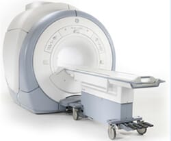
Amid the Exam: A traditional MRI unit is an extensive round and hollow magnet with a focal opening. A sliding table rests in the opening. You will lie on the restricted table and be easily situated. A little loop might be put around the region being inspected. The table will at that point be slid into the opening. The technologist will be in an abutting room, however can see, hear, and address you constantly. At times a companion or relative may remain in the live with you. Amid the exam you should stay still. A MRI exam generally comprises of a few arrangements, each enduring 2-15 minutes. Slight development between arrangements is permitted. The exam is effortless. You may feel warmth in the territory being inspected.
"Short bore" frameworks are shorter and more extensive and don't absolutely encase the patient. "Open" MRI frameworks are accessible for the individuals who can't utilize a traditional MRI; however the picture quality shifts.
Time Required: 30 to 45 minutes