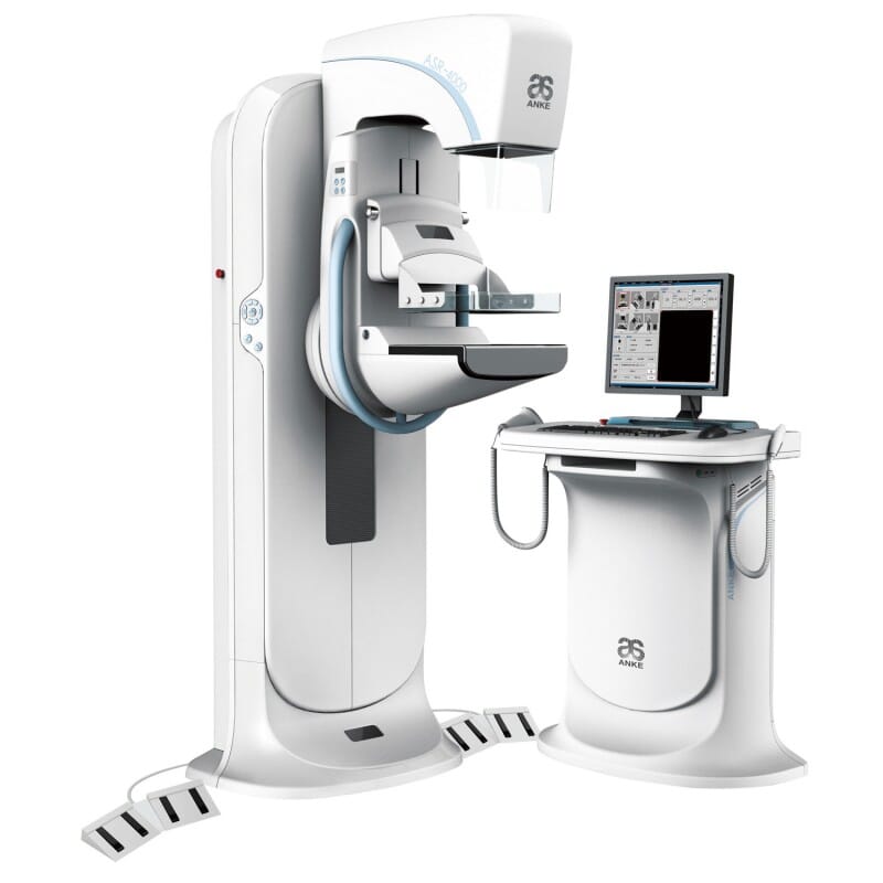
Digital Mammography test or Mammogram In Hyderabad
Mammography is specific therapeutic imaging that uses a low-measurements x-beam framework to see inside the bosoms. A mammography exam, called a mammogram, helps in the early identification and conclusion of bosom infections in ladies.
A x-ray (radiograph) is a noninvasive medicinal test that enables doctors to analyze and treat therapeutic conditions. Imaging with x-beams includes uncovering a piece of the body to a little measurement of ionizing radiation to create photos of within the body. X-beams are the most established and most every now and again utilized type of medicinal imaging.
Three late advances in mammography incorporate computerized mammography, PC supported discovery and bosom tomosynthesis.
Advanced mammography, likewise called full-field computerized mammography (FFDM), is a mammography framework in which the x-beam film is supplanted by gadgets that change over x-beams into mammographic photos of the bosom. These frameworks are like those found in computerized cameras and their proficiency empowers better pictures with a lower radiation measurements. These pictures of the bosom are exchanged to a PC for audit by the radiologist and for long haul stockpiling. The patient's understanding amid a computerized mammogram is like having an ordinary film mammogram.
PC supported discovery (CAD) frameworks look digitized mammographic pictures for anomalous regions of thickness, mass, or calcification that may show the nearness of growth. The CAD framework features these zones on the pictures, alarming the radiologist to painstakingly survey this region.
Bosom tomosynthesis, likewise called three-dimensional (3-D) mammography and computerized bosom tomosynthesis (DBT), is a propelled type of bosom imaging where various pictures of the bosom from various points are caught and recreated ("orchestrated") into a three-dimensional picture set. Along these lines, 3-D bosom imaging is like processed tomography (CT) imaging in which a progression of thin "cuts" are gathered together to make a 3-D recreation of the body.
In spite of the fact that the radiation measurements for some bosom tomosynthesis frameworks is somewhat higher than the dose utilized as a part of standard mammography, it stays inside the FDA-affirmed safe levels for radiation from mammograms. A few frameworks have measurements fundamentally the same as customary mammography.
Extensive populace considers have demonstrated that screening with bosom tomosynthesis brings about enhanced bosom tumor recognition rates and less "call-backs," cases where ladies are gotten back to from screening for extra testing as a result of a possibly anomalous finding.
Bosom tomosynthesis may likewise bring about:
prior location of little bosom malignancies that might be covered up on a regular mammogram
more noteworthy precision in pinpointing the size, shape and area of bosom variations from the norm
less pointless biopsies or extra tests
more noteworthy probability of recognizing different bosom tumors
clearer pictures of variations from the norm inside thick bosom tissu

